Spatial Omics
Bring omics data into the spatial dimension and reconstruct molecular expression and tissue architecture in situ.
10x Visium Spatial Transcriptomics
10x Visium spatial transcriptomics is a high-throughput spatial-omics technology from 10x Genomics that overcomes the loss of spatial information in conventional single-cell transcriptomics, simultaneously acquiring whole-genome expression profiles and spatial coordinates while preserving tissue morphology—enabling 'morphology visualization + molecular quantification'. Built on gridded spatial-barcode chips compatible with fresh-frozen and FFPE tissue, it maps gene-expression spatial landscapes to resolve heterogeneity, cell–cell interactions, and microenvironment zoning. Applied in TME, developmental atlases, neuroscience circuits, and pathology.
Principle Introduction
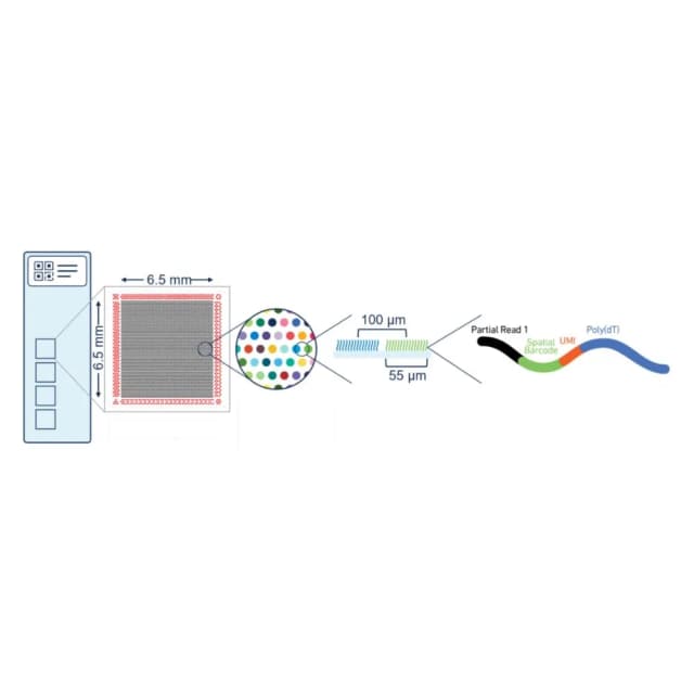
Based on 'spatial barcode labeling–in-situ capture–sequencing': tissue sections are placed on a Visium chip whose millions of spots carry unique spatial barcodes and capture probes at fixed coordinates. Permeabilization releases mRNA that binds neighboring probes, tagging each molecule with its spot barcode. After poly(A) capture, RT, amplification, and library prep, sequencing reads map back to coordinates via spatial barcodes and overlay with H&E to generate a 3-in-1 'morphology–expression–space' map.
Literature
Published in Nature Genetics, 'Multi-omic profiling highlights factors associated with resistance to immuno-chemotherapy in non-small cell lung cancer' collected pre/post neoadjuvant immunotherapy tumors from 19 NSCLC patients, mapping spatial expression via Visium and inferring cellular composition with STRIDE to identify 15 niches. Non-responders showed enriched COL11A1⁺ cancer-associated fibroblasts at tumor boundaries, spatially anti-correlated with CD8⁺ T cells; CAF formed a physical barrier via DDR1-collagen and co-localized with SPP1⁺ macrophages, worsening immune suppression.
Sample Requirements
5–10 μm sections, ≥3/sample on charged slides; -80°C sealed, dry ice; freeze within 30 min.
paraffin block or 5–10 μm sections, ≥3/sample; block at ambient, sections sealed refrigerated; note fixation/embedding/pathology.
≥0.2×0.2×0.1 cm, freeze/fix promptly; note type, grade, history; include core/boundary/adjacent.
fresh snap-frozen, 5 μm, -80°C dry ice; FFPE fixed strictly.
500 μm–1 mm, ≥5, 4°C or frozen; note culture, induction.
Deliverables
Raw data
Fastq, spatial barcode stats, QC (Q30, GC), spot signal.
Analysis
spatial expression map, regional differential genes (volcano/heatmap), cell-type spatial inference, pathway enrichment, H&E overlay.
FAQ
Cause signal loss; use charged slides, control temperature, optimize dewaxing, handle gently.
Deconvolution (STRIDE, CARD) maps cell types to spots; label transfer refines annotation.
Uniform thickness, optimize permeabilization, good RNA, increase depth.
