
Protein Expression & Detection
From high-sensitivity detection to spatial localization — a complete solution for precisely quantifying target protein expression.
Dual-Luciferase Reporter Assay
The dual-luciferase reporter assay (Dual-LUC) uses luciferase-catalyzed luminescence to study gene-expression regulation (promoter activity, transcription-factor binding, signaling). By introducing two luciferases (report and internal control), it overcomes sample and transfection-efficiency variation of single-luciferase assays for precise quantification. With high specificity, sensitivity, and reproducibility, it dynamically reflects regulation and is widely used in promoter analysis, TF–DNA validation, miRNA-target verification, and pathway activity. Unlike EMSA or Y1H, Dual-LUC mimics physiological regulation in living cells, making it essential for mechanism studies.
Principle Introduction
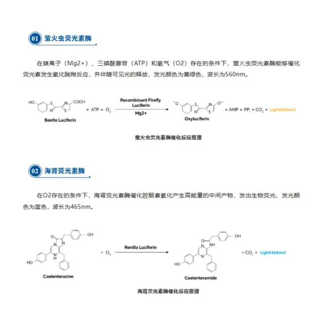
Dual-LUC uses Firefly (FLuc) and Renilla (RLuc) luciferases from different sources; detecting both enables detailed, accurate analysis. Build a reporter vector by cloning the promoter/regulatory fragment downstream of FLuc (e.g., pGL4.10); an RLuc internal-control vector (e.g., pRL-TK) is unaffected by the target. Co-transfect into cells with empty and mutant-promoter negative controls. After lysis, add substrates: FLuc catalyzes D-luciferin (yellow-green, ~560nm), RLuc catalyzes coelenterazine (blue, ~480nm). Normalize FLuc by RLuc (Fluc/Rluc ratio) to remove transfection/viability differences and reflect true regulatory level.
Literature
Journal of Neuroinflammation published "Exosomal miR-409-3p secreted from activated mast cells promotes microglial migration, activation and neuroinflammation by targeting Nr4a2 to activate the NF-κB pathway." Dual-LUC confirmed miR-409-3p's role: bioinformatic prediction of Nr4a2 3'UTR sites led to WT and seed-mutant (MUT) reporter constructs. Co-transfecting these with miR-409-3p mimic (or negative control) and Renilla internal control, dual-LUC showed the mimic strongly repressed WT but not MUT activity—providing definitive evidence that Nr4a2 is a direct miR-409-3p target.
Sample Requirements
≥500ng/μL, ≥50μL, -20℃, cold pack; attach sequencing and map.
cryopreserved cells viability >90%, dry ice; fresh protoplasts, cold pack, within 24h; note type and culture.
purified PCR fragment (≥200ng/μL, ≥30μL) or FASTA, -20℃, cold pack.
>1g/sample, snap-frozen -80℃, dry ice; specify type (leaf, callus).
Deliverables
Transfection and detection raw data
Fluc, Rluc, Fluc/Rluc tables; raw signal plots.
Quantification report
statistics (t-test/ANOVA), significance, relative-activity bar charts.
Technical documentation
optimization tips (transfection, ratio), troubleshooting, follow-up design (EMSA, ChIP).
FAQ
① wrong vector (re-sequence orientation/CDS); ② imbalanced ratios (typically 10:1:5); ③ wrong cell type; ④ needs signal induction.
① more pRL-TK; ② stronger promoter (pRL-CMV); ③ incomplete lysis; ④ expired substrate.
① fresh substrate; ② no RNase/protease in lysis; ③ read within 30 min; ④ use signal-stabilizing luminometer.
