
Protein Expression & Detection
From high-sensitivity detection to spatial localization — a complete solution for precisely quantifying target protein expression.
EMSA
Electrophoretic mobility shift assay (EMSA), also gel shift, is a classic in vitro technique detecting protein–nucleic-acid (DNA/RNA) interactions. It complements in vivo methods (e.g., ChIP) that cannot directly show binding affinity, precisely identifying DNA-binding proteins, binding specificity/affinity, mutation effects, and binding-site localization. Simpler, cheaper, and more intuitive than ChIP-seq, EMSA is the "gold standard" for verifying direct TF–promoter binding, widely used in plant development, animal disease, and regulatory networks.
Principle Introduction
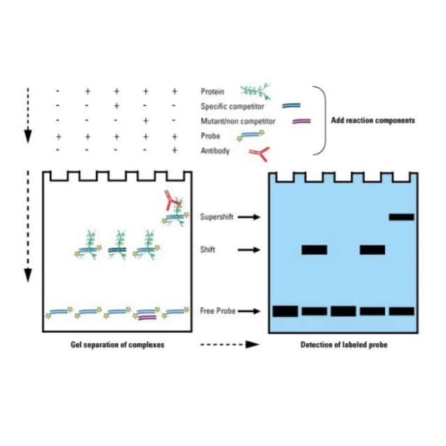
EMSA exploits that a protein bound to a labeled nucleic-acid probe forms a larger, more complex complex migrating slower than free probe in non-denaturing PAGE. Protein (P) and site (D) reach equilibrium P+D⇌PD; when association exceeds dissociation, PD is stable. After electrophoresis, free probe migrates fast while PD lags as a "shifted band"; multiple sites yield multiple bands. Competition probes or antibodies further verify specificity and identify the bound protein.
Literature
Plant Physiology published "Transcription factor CrWRKY42 coregulates chlorophyll degradation and carotenoid biosynthesis in citrus," using EMSA to clarify fruit coloration. From a color mutant they identified CrWRKY42 and designed biotin-labeled probes for W-boxes in CrBCH1, CrLCYB2, CrNYC, CrSGR promoters. EMSA showed clear shifts; excess unlabeled probe weakened the shift, mutant W-box probe gave none. Supershift with anti-CrWRKY42 antibody confirmed specificity. Combined with Y1H and dual-luciferase, CrWRKY42 binds multiple pathway promoters, coordinately driving chlorophyll degradation and carotenoid accumulation for green-to-yellow transition.
Sample Requirements
≥0.5mg/mL, ≥200μL, purity >80%, -80℃, dry ice, avoid freeze–thaw.
fresh leaf/fruit >1g, snap-frozen -80℃, dry ice; specify tissue and condition.
tissue >0.5g or cell pellet >1×10^7, snap-frozen -80℃, dry ice; provide source.
dry or solution, ≥10μM, ≥50μL, -20℃ dark, cold pack; attach sequence and label type.
Deliverables
EMSA raw data
gel images, transfer films, band grayscale quantification.
Specificity report
specific/non-specific competition and supershift results with shift calls.
Technical documentation
probe-design tips, binding-condition table, troubleshooting.
FAQ
add Poly(dI-dC), raise salt (100–200mM NaCl), lower protein, shorten probe to core site.
use 4–6% gel, soak in transfer buffer 10–15 min, keep apparatus stable.
more/stronger antibody, different epitope, longer incubation, or optimize binding first.
