Protein Expression & Detection
From high-sensitivity detection to spatial localization — a complete solution for precisely quantifying target protein expression.
BiFC Assay
Bimolecular fluorescence complementation (BiFC) visualizes protein interactions and subcellular localization in living cells via fluorescent-protein fragment reconstitution. It overcomes in vitro assays' inability to reflect the cellular environment, validating interactions in protoplasts, tissues, or whole plants with high intuitiveness, specificity, no substrate need, and dynamic tracking. BiFC reveals interaction loci (nucleus, cytoplasm, membrane) and, unlike Y2H, needs no reporter activation, more faithfully mimicking native conditions—an indispensable tool for in-cell interaction studies.
Principle Introduction
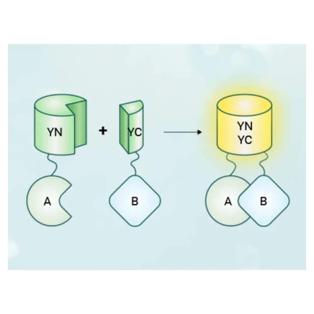
BiFC uses split fluorescent proteins. Taking Venus: the N-fragment (YN, aa 1–172) and C-fragment (YC, aa 155–238) are non-fluorescent alone. Fusing YN to protein A (A-YN) and YC to protein B (B-YC) and co-expressing in live cells, interaction brings fragments within ~10 nm; under proper conditions they refold into native-like Venus and emit yellow fluorescence. Microscopy reveals signal presence and intracellular position, judging interaction and its subcellular region.
Literature
Plant Biotechnology Journal published "A fungal CFEM-containing effector targets NPR1 regulator NIMIN2 to suppress plant immunity." Y2H screening of 11 candidates showed MdNIMIN2 strongly interacts with CfEC12. For in vivo validation, CfEC12-NYFP and MdNIMIN2-CYFP were agro-infiltrated into tobacco leaves (1:1). After 48h, confocal microscopy showed clear YFP signal in epidermal nuclei, absent in negative controls—directly proving specific interaction within plant nuclei and laying the foundation for elucidating CfEC12's immune-suppression via MdNIMIN2.
Sample Requirements
≥500ng/μL, ≥50μL, -20℃, cold pack; attach sequencing and map.
fresh leaf/callus >1g/sample, 4℃ cold transport, within 24h; note variety and state.
cryopreserved cells viability >90%, dry ice; fresh protoplasts, cold pack, within 12h; note type and culture.
bait/prey sequences (FASTA), specify restriction sites and fusion orientation.
Deliverables
Fluorescence raw data
confocal photos of test/control, signal-intensity quantification, subcellular-localization maps.
Interaction report
conclusion on interaction and subcellular locus from signal distribution.
Technical documentation
optimization tips (fragment choice, transformation), troubleshooting, follow-up design (Co-IP, Pull-Down).
FAQ
① self-associating fragment (improved Venus); ② fusion error (re-sequence); ③ autofluorescence (lower-background material or mCherry); ④ lower expression.
① better fragment pair (SPYNE/SPYCE, YN155/YC155); ② more vector; ③ enhancer or longer exposure; ④ more sensitive confocal.
① in vitro BiFC with purified fusions; ② truncation mapping; ③ combine in vitro Pull-down.
