
Exosomics
Harness the tiny messengers of cell-to-cell communication to advance early disease screening and dissect precise mechanisms of action.
Exosomal Metabolomics Analysis
Exosome Metabolomics (EM) is a systematic qualitative, quantitative, and functional analysis of small-molecule metabolites in exosomes (typically <1500 Da), covering amino acids, organic acids, sugars, lipids, nucleotides, vitamins, etc. Vesicle-membrane-protected exosomal metabolites resist enzymatic degradation and environmental interference in biofluids, are highly stable in serum/urine, and faithfully reflect the source cell's metabolic reprogramming. Versus routine cell/tissue metabolomics, EM offers three advantages: ① non-invasive samples—biofluids, no invasive procedures, ideal for clinical monitoring and large cohorts; ② functional targeting—exosomal metabolites are actively sorted functional molecules participating in intercellular metabolic communication; ③ low interference—purification removes high-abundance free metabolites, giving higher specificity. EM is now core for disease metabolic mechanisms, non-invasive biomarker development, drug-target screening, and precision delivery-carrier design, widely used in cancer, neurodegeneration, and metabolic syndrome.
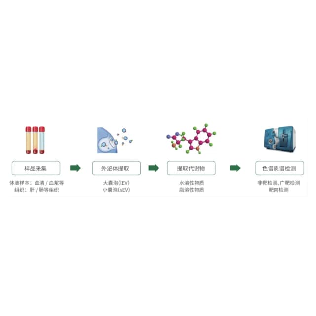
Principle Introduction
Exosome isolation and purity validation via efficient purification; metabolite extraction and pretreatment—low-temperature lysis + efficient extraction, lysis, cleanup, normalization, with concentration corrected by exosomal protein content; high-resolution MS detection—mainly Thermo Orbitrap platforms for sensitivity and accurate identification in non-targeted mode; targeted validation—MRM quantification of differential metabolites; data analysis and functional interpretation—database-driven standardized pipeline for identification/quantification and matching to authoritative databases to determine metabolite class and relative/absolute content; differential analysis—screen disease vs healthy metabolites (|FC|≥1.2, P<0.05); functional enrichment—KEGG pathway analysis interpreted with clinical phenotypes.
Literature
In ACS Publications, "Proteomic, Metabolomic, and Fatty Acid Profiling of Small Extracellular Vesicles from Glioblastoma Stem-Like Cells and Their Role in Tumor Heterogeneity." performed metabolomics on glioblastoma stem cell (GSC)-derived exosomes, focusing on amino-acid and fatty-acid features. GSC exosomes are enriched in amino-acid/organic-acid transport-related metabolites that nourish neighboring tumor cells; saturated fatty acids are high in GSC exosomes whereas the parent cells are dominated by unsaturated fatty acids, indicating highly regulated metabolite sorting; different GSC subtypes (mesenchymal/proneural) show specific exosomal metabolite differences mediating intercellular molecular cooperation. This first revealed the core role of exosomal metabolites in tumor heterogeneity, offering new directions for glioblastoma metabolic targeting.
Sample Requirements
serum/plasma ≥0.5 mL; urine ≥5 mL; CSF ≥1 mL; saliva ≥3 mL.
≥10 mL.
≥1×10¹⁰.
total ≥20 μg.
Deliverables
QC report
exosome purity (NTA, Western blot, TEM); metabolite quality (extraction efficiency, MS quality); quantitation quality (non-targeted CV ≤15%, targeted recovery 90%–110%).
Metabolite quantification and annotation
matrix (name, formula, MW, relative/absolute content, FC, P); differential analysis (volcano/heatmap with significant metabolites flagged); database annotation (HMDB, METLIN, KEGG; function and pathway).
Functional analysis
KEGG enrichment plots (Top20, e.g., glycolysis, fatty-acid β-oxidation), metabolite pathway network; clinical association (ROC of differential metabolites vs diagnosis/prognosis).
Report
conclusions (core differential metabolite list, biomarker candidates); interpretation of regulatory mechanisms and exosome source inference.
FAQ
By study stage: ① non-targeted—exploratory, unbiased, discovers new biomarkers, slightly lower quantitative precision; ② targeted—confirmatory, high sensitivity, suited to large clinical validation, requires known metabolites. Recommended: non-targeted screening → targeted validation.
Plasma is better: ① less interference—serum clotting-factor activation releases free metabolites that dilute exosomal ones; EDTA plasma avoids coagulation and yields higher exosomal-metabolite proportion; ② stability—EDTA inhibits metalloproteases, degradation (<5%) lower than serum (>15%). Notes: avoid heparin; serum needs clot removal, plasma only needs platelet removal.
Yes—via high-sensitivity techniques and optimized workflow: ① minimum 0.2 mL serum / 3 mL urine; ② nano-extraction columns raising recovery >85%; ③ Orbitrap Exploris 120 with 10 scans/s to boost low-abundance signals; ④ locally weighted regression baseline correction to reduce false positives. Example: successfully profiled 1 mL CSF exosomes from a rare-disease patient, identifying 320 metabolites with valid differential analysis.
