
Exosomics
Harness the tiny messengers of cell-to-cell communication to advance early disease screening and dissect precise mechanisms of action.
Exosomal Proteomics Analysis
Exosome quantitative proteomics is a high-throughput technique for the systematic separation, identification, and precise quantification of the exosomal proteome. As cell-secreted membrane vesicles, exosomes carry proteins that are core functional mediators of intercellular communication; protected by the vesicle membrane, they are highly stable in biofluids and faithfully reflect the source cell's status. Versus routine cell/tissue quantitative proteomics, exosome quantitative proteomics focuses on exosomes as a "functional carrier," uses non-invasive fluids, and better suits clinical monitoring and large cohorts; versus qualitative exosome proteomics, it uses labeled or label-free quantification to precisely measure protein differences, avoiding the limits of pure identification and better serving biomarker screening and mechanism validation.
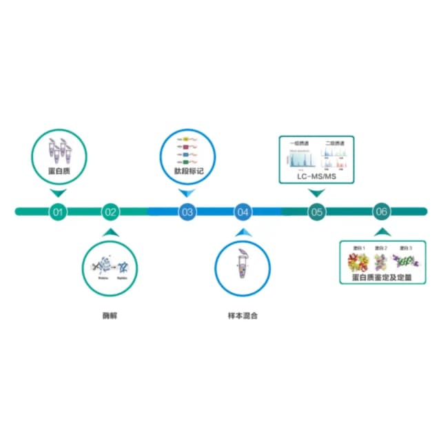
Principle Introduction
Core principle: exosome purification, protein enrichment/quantification, mass-spectrometry detection, functional analysis. Isolate and verify exosomes (remove debris/free protein); extract and preprocess exosomal proteins; digest proteins into 8–20 aa peptides; desalt via C18 SPE to remove impurities/salts and concentrate peptides; labeled or label-free quantification; MS detection on a high-resolution platform in "MS1 quantification, MS2 identification" mode to obtain peptide m/z, intensity, and sequence, ensuring accurate identification and quantification; bioinformatics aligns MS data to the human protein database to select high-confidence proteins, derives a quantification matrix, and mines core functions via GO/KEGG enrichment and PPI networks.
Literature
In J Cachexia Sarcopenia Muscle, "Proteomic profiling of human plasma extracellular vesicles identifies PF4 and C1R as novel biomarker in sarcopenia" collected plasma exosomes from healthy controls and sarcopenia patients (>65 years) and used proteomics plus bioinformatics to identify novel sarcopenia biomarkers. This was the first proteomic analysis of plasma exosomes in sarcopenia. Data showed that patient exosomes carry proteins related to calcium/collagen metabolism, immune response, and inflammation. PF4 and C1R emerged as promising diagnostic biomarkers usable in routine clinical practice to identify sarcopenia and enable earlier prevention and intervention.
Sample Requirements
serum ≥5 mL; plasma ≥5 mL; urine ≥15 mL; CSF ≥2 mL; saliva ≥5 mL.
≥30 mL.
≥300 μL.
total ≥50 μg, concentration ≥1 μg/μL.
Deliverables
QC report
exosome purity (NTA histogram, Western blot markers, TEM morphology); protein/peptide quality (extraction yield, desalting, SDS-PAGE, MS quality); quantitation quality (isobaric tag balance for labeled, peptide peak-area reproducibility for label-free).
Protein quantification and annotation
quantification matrix (relative values, FC, P, FDR); differential protein screening (volcano/heatmap with name, MW, pI, trend); database annotation (function, subcellular localization, domains, disease association).
Functional analysis
GO/KEGG enrichment plots (Top20), PPI network, differential-protein clustering heatmap.
Report
conclusions (core differential protein set, biomarker candidates, association strength with disease/phenotype).
FAQ
Yes—hemolysis releases hemoglobin interfering with extraction/MS; lipemia (high triglycerides/cholesterol) impairs exosome isolation and protein purity. Handle by: avoiding vigorous shaking, prompt centrifugation, and strict exclusion of hemolyzed/lipemic samples; if unrecollectable, use a hemoglobin-removal kit for hemolysis or ultracentrifugation to remove the lipid layer for lipemia, then isolate exosomes.
Three steps: ① lab validation—Western blot/ELISA on a large cohort (≥200) to confirm diagnostic/prognostic association; ② functional validation—cell experiments for biological function; ③ clinical translation—develop a rapid assay and validate sensitivity/specificity/stability in multi-center samples before clinical use.
