
Transcriptomics
Capture gene-expression dynamics with precision and reveal regulatory mechanisms — reconstructing gene-response patterns under any experimental condition.
MicroRNA (miRNA) Sequencing
MicroRNA sequencing (miR-seq) is a high-throughput technique for the ~18–25 nt non-coding small RNA (miRNA), enabling systematic quantification of known miRNAs, discovery of novel miRNAs, and analysis of sequence variants and base modifications (e.g., m⁶A) at single-base resolution with whole-genome coverage and high sensitivity. Versus qPCR, miR-seq unbiasedly detects low-abundance miRNAs and species-specific novel miRNAs/variants; versus microarrays, it offers a wider dynamic range and more faithful reflection of expression differences. As core regulators, miRNAs drive proliferation, differentiation, and apoptosis via target mRNA degradation or translational repression, and their dysregulation is linked to cancer, neurodegeneration, and metabolic syndrome—making miR-seq key for biomarker screening, mechanism studies, and drug-target discovery.
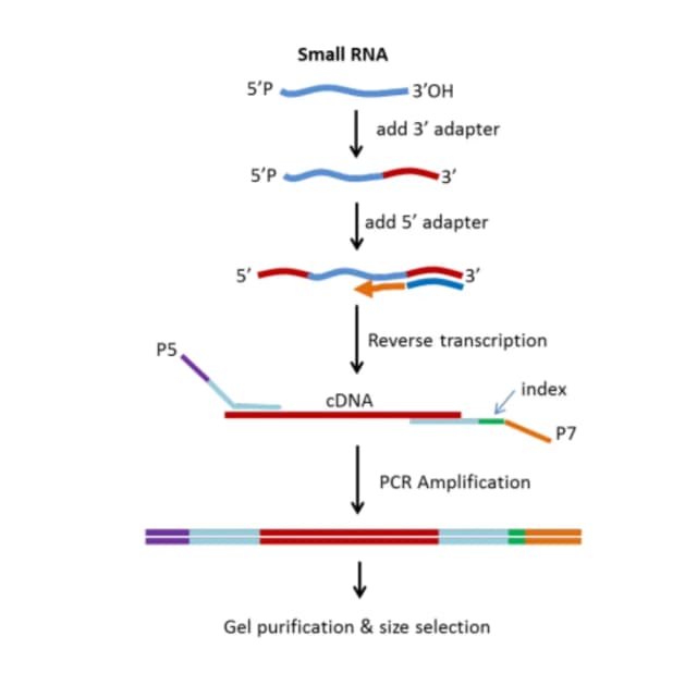
Principle Introduction
miRNA sequencing: sample processing and small-RNA enrichment (RNA extraction, QC); dedicated library construction (adapter design, reverse transcription and amplification, strand-specific retention) on Illumina NovaSeq 6000/MiSeq (Q30 ≥85%); data analysis—filter low-quality reads, adapters, and non-miRNA fragments (rRNA/tRNA); miRNA identification by aligning to miRBase and quantifying, novel miRNA prediction with miRDeep2/miREvo plus secondary-structure validation; variant analysis (isomiRs, editing sites); functional annotation via TargetScan/miRanda target prediction and GO/KEGG enrichment and miRNA–mRNA co-expression networks.
Literature
In iScience, "Direct microRNA sequencing using Nanopore Induced Phase-shift Sequencing (NIPSS)" addressed the inability of conventional miR-seq to preserve modifications by developing nanopore phase-shift sequencing (NIPSS), the first single-molecule direct miRNA sequencing. It distinguished the highly similar miR-21 (oncomiR) from miR-21+U (3′ mono-U variant) and from let-7a (tumor suppressor) at single-base resolution; introduced m⁶A into miR-21 and located the modification via NIPSS signal differences—the first single-molecule direct detection of miRNA modification; 15-nt reads discriminated 99.5% of human miRNAs, providing a new tool for studying miRNA modification abnormalities in cancer.
Sample Requirements
≥100 mg; low-RNA tissues (fat/skin/bone) ≥1 g.
tender leaf/stem/flower/fruit ≥500 mg; seeds ≥2 g.
≥10⁶.
fresh ≥5 mL.
≥2 tubes.
≥10⁷ / 300 mg.
total ≥2 μg, concentration ≥100 ng/μL, volume ≥20 μL.
Deliverables
QC
RNA quality, sequencing quality, filtering results.
miRNA identification and quantification
known miRNA expression matrix (RPM/TPM), differential miRNA list (log2FC, P, FDR), volcano/heatmap; novel miRNA predicted sequences (FASTA), secondary-structure diagrams, expression; isomiR types and proportions (Illumina), m⁶A and other modification sites/signal (NIPSS).
Functional analysis
target prediction (TargetScan/miRanda tables); GO/KEGG enrichment scatter plots, miRNA–mRNA co-expression networks; database annotation (miRBase ID, target function via Swiss-Prot/KEGG, disease association via OMIM).
Report
core differential miRNA set, biomarker candidates, key pathways; expression trends vs disease/treatment and biological meaning of modifications/variants.
FAQ
Prefer EDTA or sodium citrate; avoid heparin—heparin binds phenols, lowering purity (OD260/230 <1.5) and inhibiting reverse transcriptase; EDTA/citrate preserve purity and RNase inhibition (<<5% degradation at 4°C/24 h). If heparin was used, digest with heparinase or recollect.
Use prediction + validation: 1) bioinformatics—TargetScan, miRanda, PITA, intersect for reliability; 2) correlation—paired miR-seq and mRNA-seq to find negative correlations; 3) validation—dual-luciferase reporter, transfection.
Sampling—snap-freeze in liquid nitrogen, or store in 2% PVP RNAlater; extraction—plant RNA kit with β-mercaptoethanol, chloroform/isoamyl alcohol (24:1) spin to remove protein/polyphenol, LiCl (2 M) precipitation to remove polysaccharides; purification—Sevag (4:1) or RNA cleanup columns if OD260/230 <1.5; QC—clear small-RNA peak (25–200 nt) on Agilent 2100 without smearing.
