
Metabolomics
Capture the direct signals of phenotypic change through precise metabolite quantification — a direct readout of physiological status.
Bile Acid Metabolomics
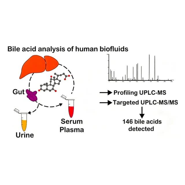
Bile acids (BAs) are steroidal compounds synthesized from cholesterol in the liver—primary BAs (CA, CDCA) and secondary BAs (DCA, LCA) from gut microbiota—and are the core functional components of bile. As "metabolic hub molecules," they both emulsify fats for absorption of lipids and fat-soluble vitamins (A, D, E, K) and act as signaling molecules activating FXR and TGR5 to regulate glucolipid metabolism, inflammation and microbiota balance. Their dysregulation links to NAFLD, gallstones, cirrhosis, diabetes and colorectal cancer, and they also matter in plant stress and aquaculture lipid metabolism. Bile-acid profiles more precisely reflect liver function, microbiota homeostasis and systemic metabolism than traditional markers, making BA testing core to diagnosis, drug development and basic research.
Principle Introduction
BA detection rests on "separation–identification–quantification" plus their physicochemical and signaling properties: 1. Separation—UPLC or LC-MS/MS exploits polarity/hydrophobicity differences among primary/secondary and conjugated/free subtypes; 2. Identification & quantification—MS matches characteristic precursor–product ion pairs for precise subtype ID, with isotope internal standards (e.g., cholic-d4, CDCA-d4) correcting matrix effects; 3. Functional association—BA-profile composition and changes are linked to signaling pathways to analyze liver metabolism, microbiota and disease.
Literature
In "Exploring serum bile acids as potential noninvasive biomarkers for nonalcoholic fatty liver disease" (Egyptian Liver Journal), serum BA profiles were analyzed to find noninvasive NAFLD biomarkers. Subjects (healthy controls, NAFLD suspects) were confirmed by liver biopsy. LC-MS/MS quantified 31 BA subtypes; mechanism showed CYP7A1/CYP27A1 abnormalities raised primary BA synthesis while microbiota imbalance (lower Bifidobacterium, higher Clostridia) altered secondary BA metabolism, disturbing the serum BA profile. Serum BA profiles are a potential noninvasive diagnostic panel, avoiding invasive biopsy.
Sample Requirements
≥500 μL, EDTA/heparin anticoagulated (plasma), −80°C sealed, dry ice; note fasting/postprandial
≥1 g, snap-freeze −80°C, dry ice; note site/storage
≥200 μL, −80°C dark sealed, dry ice; note source (hepatic/gallbladder)
plant ≥2 g, animal ≥1 g, cell pellet ≥1×10⁷, −80°C, dry ice; note site/treatment
≥1 mL, −80°C sealed, dry ice; 0.1% NaN₃ optional, note 24 h/morning
Deliverables
Raw data
LC-MS/MS raw chromatograms, BA-subtype retention times, characteristic-ion match reports, peak-area raw data.
Data analysis
BA-subtype identification, quantitative table (ng/mL or μg/g), composition-ratio plots, differential vs control/library.
FAQ
Add SPE/liquid–liquid cleanup, optimize chromatography (pH, column), use MRM to avoid interference, use higher-purity standards/reagents.
Inconsistent collection/handling or instrument drift—standardize, calibrate, run parallel samples (RSD≤10%).
