Proteomics
Dissect the proteome to uncover key biomarkers and decode the mechanisms of life from the terminal executors of function.
4D-DIA Proteomics
4D-DIA proteomics fuses 4D separation (retention time, m/z, ion intensity, ion mobility) with data-independent acquisition (DIA), a next-generation high-throughput proteomics technology. Its core innovation is the added ion-mobility (IM) dimension, which improves peptide resolution via extra spatial separation and, combined with DIA's unbiased acquisition of all peptides, overcomes the limited coverage and poor low-abundance protein detection of conventional 3D-DIA (no ion mobility). 4D-DIA offers higher sensitivity (ion-mobility filtering raises low-abundance protein detection by 30–50%), more accurate quantification (peptide-specific matching cuts co-elution interference; quantitative CV ≤10%), and broader coverage (10,000+ proteins per run with stable modified-protein detection). It is a core technology for clinical samples, complex-disease biomarker screening, drug-target validation and multi-omics studies.
Principle Introduction
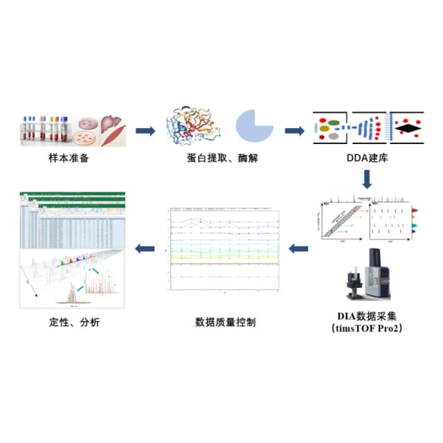
Data-independent acquisition (DIA) is a recently developed MS acquisition mode that performs unbiased MS analysis of all signals in complex samples, yielding higher peptide coverage. The full MS scan range is divided into windows; each window's ions are rapidly and cyclically selected, fragmented and detected, capturing all fragment information without omission or bias. 4D-DIA, built on the timsTOF Pro2 instrument, adds a fourth dimension—ion mobility—to the traditional 3D data (retention time, m/z, ion intensity). Using diaPASEF, it boosts protein identification, sensitivity and data completeness. Workflow: sample pretreatment and protein preparation, multi-dimensional peptide separation, 4D-DIA MS acquisition, data analysis and functional interpretation.
Literature
In "Machine learning-based analysis identifies and validates serum exosomal proteomic signatures for the diagnosis of colorectal cancer" (Cell Reports Medicine), 4D-DIA proteomics was used to profile serum exosomal protein differences between colorectal cancer patients and healthy controls, with a machine-learning diagnostic model. A total of 1,200+ exosomal proteins were identified and 10 core marker combinations selected; the model achieved AUC 0.96 for colorectal cancer and AUC 0.92 for early-stage disease, providing a highly specific, sensitive liquid-biopsy approach. The high-depth, reproducible quantification precisely captured low-abundance exosomal differences, supplying high-quality data for ML-based marker discovery.
Sample Requirements
fresh tissue ≥50 mg (animal) or ≥200 mg (plant);
5–10 μm sections ≥10 slides, tissue area ≥20 mm²;
≥1×10⁶ cells; cell slides ≥5;
serum/plasma ≥1 mL; urine ≥10 mL; CSF ≥0.5 mL, saliva ≥3 mL;
protein ≥50 μg; peptide ≥20 μg.
Deliverables
QC report
sample quality (protein yield, peptide desalting, SDS-PAGE); 4D acquisition quality (ion-mobility resolution, peptide peak capacity, MS2 count, Q30); quantification quality (protein/peptide IDs, quantitative CV, biological-replicate correlation).
Protein quantification & annotation
expression matrix (FPKM/Intensity, FC, P, FDR); differential protein list, volcano/heatmap/cluster plots; database annotation (function, subcellular localization, domains, modification sites).
Functional analysis
GO/KEGG/Reactome enrichment (Top20 pathways), PPI network, differential-protein cluster heatmap; multi-omics links (protein–metabolite/miRNA networks).
Interpretation report
key differential proteins, biomarker candidates, regulatory pathways; biological mechanisms and technical advantages.
FAQ
Yes—it is the optimal proteomics solution for FFPE: ion mobility selects intact peptides and filters degradation fragments, raising protein IDs by 30–40% vs 3D-DIA. Notes: thorough deparaffinization (xylene + ethanol), extended digestion (20 h, high-activity trypsin), SDS-PAGE verification of integrity.
timsTOF Pro 2 (IM benchmark, resolution ≥100, ≥100 Hz) for low-abundance/exosomal samples; Orbitrap Astral (≤1 ppm) for PTM and complex quantification; SCIEX ZenoTOF 7600 (≥200 Hz) for large cohorts.
Use same-source, same-batch samples; match protein trends to mRNA (mRNA unchanged but protein up suggests post-translational regulation); focus on pathways co-enriched across omics; build mRNA–protein–metabolite networks with Metascape/String.
