
Proteomics
Dissect the proteome to uncover key biomarkers and decode the mechanisms of life from the terminal executors of function.
PRM Targeted Proteomics
PRM targeted proteomics (Parallel Reaction Monitoring) is a precise targeted quantification method on high-resolution MS, focusing on specific detection and quantification of known target proteins/peptides. Predefined MS parameters enable targeted scanning to efficiently capture and quantify target proteins (and modification sites) from complex samples. It resolves the low throughput and cross-reactivity of ELISA and the limited accuracy and low-abundance detection of untargeted proteomics.
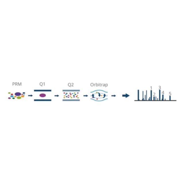
Principle Introduction
PRM is an LC-MS/MS targeted method providing selective, specific, reproducible and accurate detection of target proteins/peptides (including modified peptides) for relative or absolute quantification. Core principle: preset target peptides, targeted MS acquisition, precise quantification. Workflow: sample pretreatment and protein preparation, target-peptide selection and method setup, PRM targeted MS acquisition, data analysis and quantitative calibration.
Literature
In "Parallel-reaction monitoring revealed altered expression of a number of epitranscriptomic reader, writer, and eraser proteins accompanied with colorectal cancer metastasis" (Proteomics), PRM combined with SILAC quantified differentially expressed epitranscriptomic reader-writer-eraser (RWE) proteins in matched primary/metastatic colorectal cancer cells (SW480/SW620). Of 113 non-redundant RWE proteins, 48 were up (>1.5×) and 5 down in metastatic cells; NAT10, hnRNPC and DKC1 promoted metastasis, while DUS1L linked to tumorigenesis. PRM effectively identified metastasis-associated epitranscriptomic regulators as potential therapeutic targets.
Sample Requirements
fresh ≥50 mg (animal) or ≥200 mg (plant); FFPE 5–10 μm ≥10 slides, area ≥20 mm²;
adherent ≥2×10⁶; suspension ≥3×10⁶;
serum/plasma ≥1 mL (routine) or ≥3 mL (low-abundance); urine ≥5 mL, CSF ≥1 mL;
protein ≥50 μg (routine) or ≥100 μg (low); peptide ≥20 μg.
Deliverables
QC report
sample quality (protein yield, peptide recovery, target-peptide detection); MS quality (fragment-ion resolution, Q30, peak-shape score); quantification quality (target CV, replicate correlation, internal-standard recovery).
Protein quantification & annotation
matrix of relative abundance (peak-area ratio) or absolute concentration (pg/mL, internal-standard corrected), FC, P, FDR; differential target list (|FC|≥1.2, P<0.05), bar/heatmap, fragment-ion spectra; function, localization, disease association, modification sites.
Functional analysis
GO/KEGG enrichment; ROC and survival curves; method validation (linear range, LOD, LOQ, recovery).
Interpretation report
quantitative results, significance and phenotype association; biological significance of core targets.
FAQ
Increase starting material and remove high-abundance proteins; select high-ionization peptides with stable-isotope internal standards; raise scan counts (≥20), extend gradient (60–90 min), optimize collision energy.
Method validation (R²≥0.99, LOD, recovery 90–110%); cross-validation shows >85% agreement for high-abundance and >70% for low-abundance proteins; isotope internal standards keep absolute-quantification deviation ≤10%.
Yes—add de-modification enzyme inhibitors, choose single-modification peptides, optimize collision energy for site-specific fragments, and spike isotope-labeled modified peptides.
