
Proteomics
Dissect the proteome to uncover key biomarkers and decode the mechanisms of life from the terminal executors of function.
Malonylation Proteomics
Malonylation Proteomics is a core sub-service of quantitative modification proteomics, focusing on protein lysine malonylation (Malonylation). Leveraging high-throughput MS detection and specific enrichment (anti-Kma antibody IP), it captures and quantifies Kma modified peptides at Lys 位點, systematically identifying modification sites, screening differentially modified proteins, and deciphering the signaling pathways and biological functions of modification regulation. Widely applied to disease-mechanism research (e.g., oncology, cardiovascular, neurodegenerative diseases), drug-target discovery, biomarker screening, and multi-omics integration, it supports clinical translation and precision medicine as a key technique for revealing 'dynamic protein functional regulation'.
Principle Introduction
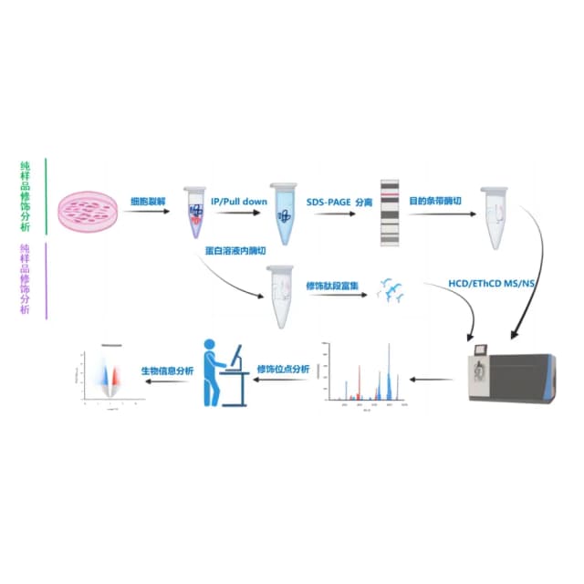
The core of Malonylation Proteomics is to enrich target modified peptides using specific antibodies or chemical-affinity reagents, combined with LC-MS/MS detection on a high-resolution MS platform (Orbitrap Astral / timsTOF Pro 2) and label-free or metabolic-labeling quantification (SILAC / TMT) strategies, achieving precise modification-site identification and inter-group differential analysis. For the chemical properties of lysine malonylation (Malonylation), a dedicated enrichment method (anti-Kma antibody IP) is used; modification-site localization probability ≥75% is classified as high confidence (Class I), with strict FDR <1% filtering, plus downstream functional analysis including GO/KEGG pathway enrichment, kinase–substrate prediction, and PPI network.
Experiment Flow
Sample pretreatment & protein preparation
lyse tissue or cells, reduce, alkylate and digest protein to peptides.
Specific enrichment of modified peptides
enrich modified peptides via TiO2/IMAC and verify enrichment efficiency.
LC-MS/MS detection
acquire spectra on a high-resolution MS platform in DIA or DDA mode.
Data analysis & functional interpretation
identify sites with FDR control, interpret by GO/KEGG/Reactome and PPI.
Modification regulation analysis
analyze signaling pathways, kinase-substrate networks and functions.
Sample Requirements
common animal tissues (liver, heart, brain, skin, kidney, intestine, muscle, various glands, etc.) ≥10 mg (risk 5 mg); for human clinical tissue samples ≥10 biological replicates recommended, common animal tissues ≥3 biological replicates;
adherent cells ≥5×10⁶, suspension cells ≥1×10⁷;
serum/plasma ≥100 μL (risk 50 μL), urine ≥1 mL (risk 0.5 mL), cerebrospinal fluid ≥1 mL;
total protein ≥100 μg, total peptide ≥50 μg;
flash-freeze in liquid nitrogen, store at -80℃ (≤3 years); aliquot into multiple tubes to avoid repeated freeze-thaw; ship on dry ice.
Deliverables
Data QC report
sample quality (protein extraction yield, peptide recovery ≥80%, modified-peptide enrichment efficiency ≥50%); MS detection quality (number of modified sites identified, peptide matching score, Q30 ratio); quantification quality (modification-site CV, biological-replicate correlation, differential-modification-site screening criteria);
Modification quantification & annotation
quantification matrix (expression level, FC, P value, FDR of modified sites); differential analysis results (differential modified-site list, volcano plot, heatmap clustered by function/pathway); database annotation (site amino-acid type, sequence position, protein function description, known modification–function associations);
Functional analysis results
GO/KEGG/Reactome pathway-enrichment scatter plot (Top 20 pathways), STRING database PPI network (highlighting core differential modified proteins and interactions), modification-regulation analysis (kinase–substrate relationship prediction);
Analysis & interpretation report
core differential modified-site set, potential biomarker/target candidates, association strength with disease/phenotype; interpretation of core-site functional mechanisms and clinical/research significance.
FAQ
Improve detection through sample pretreatment, enrichment optimization, and MS parameter tuning: ①Sample level: increase starting sample amount and use high-activity digestion reagents to extend digestion time; ②Enrichment level: adopt pre-enrichment + secondary enrichment to reduce non-specific peptide interference; ③MS level: increase sequencing depth, use high-resolution MS, and extend gradient elution time.
Quantification accuracy is ensured by technical design and experimental validation. ①Technical: add isotope-labeled internal standards during enrichment to correct enrichment-efficiency bias; MS quantification uses "peptide-intensity normalization" to ensure inter-group comparability; ②Experimental: select 10–20 differential modification sites for PRM validation, achieving >80% consistency.
Screen functional modification sites through multi-dimensional validation. ①Conservation analysis: use databases such as PhosphoSitePlus and UniProt to verify conservation of the modification site across homologous proteins; ②Functional association: check whether the site has been reported or overlaps with a known functional domain; ③Experimental validation: introduce site mutations or overexpress the modifying enzyme and measure changes in protein activity and cellular function to verify the modification's functional impact.
